Home
Uncategories
Back Of Skull Anatomy - Stock Illustration - Human skull, back view. Clipart gg59791552 - GoGraph / All the bones of skull, joined together by sutures, are immobile and create the cranium, with the exception.
Back Of Skull Anatomy - Stock Illustration - Human skull, back view. Clipart gg59791552 - GoGraph / All the bones of skull, joined together by sutures, are immobile and create the cranium, with the exception.
Back Of Skull Anatomy - Stock Illustration - Human skull, back view. Clipart gg59791552 - GoGraph / All the bones of skull, joined together by sutures, are immobile and create the cranium, with the exception.. Related posts of bone of back of skull. 12 photos of the bone of back of skull. A thorough description is beyond the. All the bones of skull, joined together by sutures, are immobile and create the cranium, with the exception. It is believed that trepanation was used to either relieve painful headaches, or to release demons from the skull.
Skull, skeletal framework of the head of vertebrates, composed of bones or cartilage, which form a unit that protects the brain and some sense organs. This anatomic region is complex and poses surgical challenges for otolaryngologists and neurosurgeons alike. Anatomical structures of the skull include: The upper back is a complex area containing a number of muscles that perform various actions on the scapulae shoulder blades and humerus. The skull base is the inferior portion of the neurocranium.
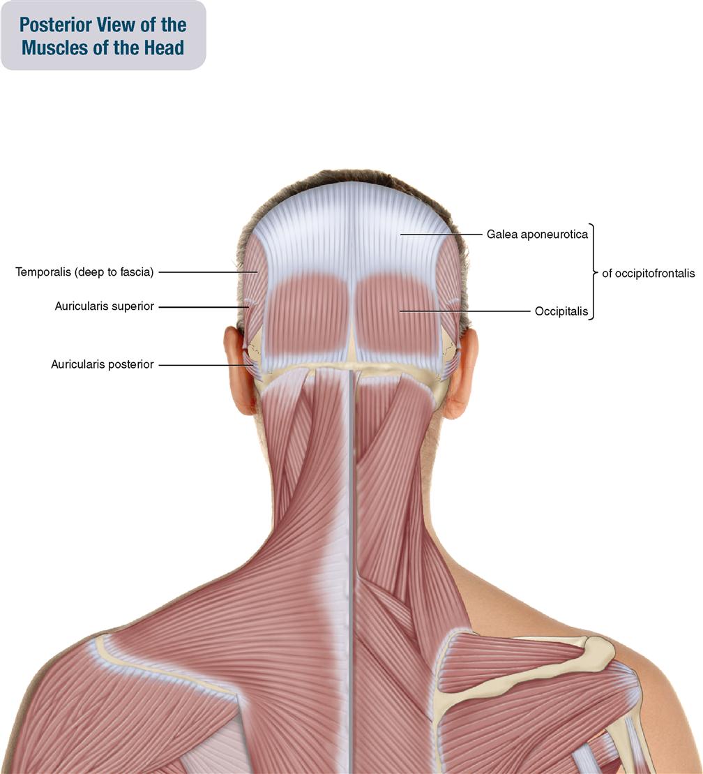
9. Muscles of the Head | Musculoskeletal Key from musculoskeletalkey.com The major sutures are the coronal suture, sagittal suture, lambdoid suture and squamosal sutures. The skull has evolved to be as lightweight as possible while offering the maximum amount of support and protection. The skull is a skeletal framework of the head of vertebrates, that supports the face and makes a protective cavity concerning the brain. The skull performs vital functions. Learn about the anatomy of the skull bones and sutures as seen on ct images of the brain. The skull includes the upper jaw and the cranium. Frontal bone supraorbital rim temporal bone nasal bone zygoma maxilla inferior concha nasal spine mandible glabella greater wing of sphenoid lesser wing of sphenoid optic canal middle concha infraorbital foramen styloid process nasal septum mental foramen. Norma basalis ( anterior part , middle part and posterior part ).
So, the human skull consists of 23 bones.
So, the human skull consists of 23 bones. Looking at it from the inside it can be subdivided into. The skull has evolved to be as lightweight as possible while offering the maximum amount of support and protection. The bbc is not responsible for the content of external websites. Learn about skull base anatomy with free interactive flashcards. This anatomic region is complex and poses surgical challenges for otolaryngologists and neurosurgeons alike. The cranium (skull) is the skeletal structure of the head that supports the face and protects the brain. It supports and protects the face and the brain. Human skull from the front. A cartilaginous mould begins to grow this is why raising your eyebrows can create the appearance that the back of the head is moving. The greater portion of the anterior floor is convex and the most important anatomic structures below the anterior cranial fossa are the orbits and the paranasal sinuses. Learn about the anatomy of the skull bones and sutures as seen on ct images of the brain. The base of the skull (or skull base) forms the floor of the cranial cavity and separates the brain from the structures of the neck and face.
The skull base is the inferior portion of the neurocranium. So, the human skull consists of 23 bones. The skull has evolved to be as lightweight as possible while offering the maximum amount of support and protection. The skull has a single occipital condyle.7 the skull consists of five major bones: The cranium (skull) is the skeletal structure of the head that supports the face and protects the brain.
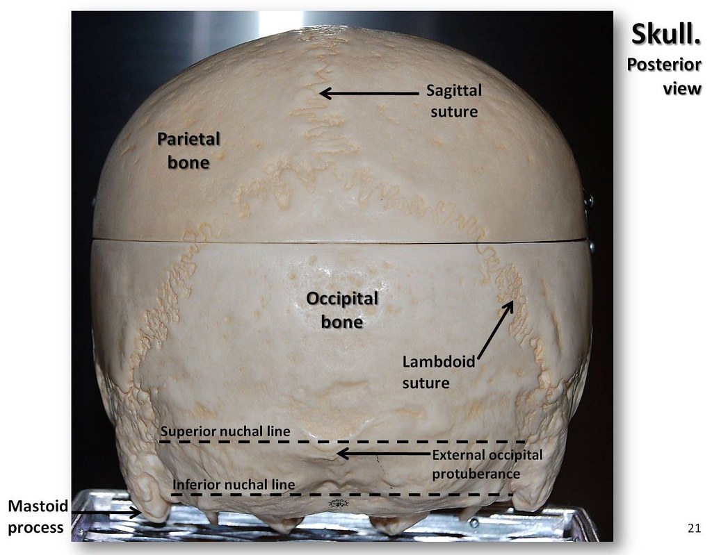
Skull, posterior view with labels - Axial Skeleton Visual … | Flickr from c1.staticflickr.com Skull, skeletal framework of the head of vertebrates, composed of bones or cartilage, which form a unit that protects the brain and some sense organs. This anatomic region is complex and poses surgical challenges for otolaryngologists and neurosurgeons alike. Skull reshaping is done on any of the structures that lie above the face. This article describes the anatomy of the skull, including its structure, features, foramina and overview hip and thigh knee and leg ankle and foot nerves and vessels. It is believed that trepanation was used to either relieve painful headaches, or to release demons from the skull. It supports and protects the face and the brain. The upper back is a complex area containing a number of muscles that perform various actions on the scapulae shoulder blades and humerus. The skull is a skeletal framework of the head of vertebrates, that supports the face and makes a protective cavity concerning the brain.
The skull or known as the cranium in the medical world is a bone structure of the head.
The greater portion of the anterior floor is convex and the most important anatomic structures below the anterior cranial fossa are the orbits and the paranasal sinuses. Skull anatomy | with labels. All the bones of skull, joined together by sutures, are immobile and create the cranium, with the exception. Continue scrolling to read more below. William is a final year medical student in australia who has taught anatomy to tertiary science and. The anterior fossa is formed by the orbital plates of the frontal bone, cribriform plate of the ethmoid, and lesser wings of the sphenoid. Frontal bone supraorbital rim temporal bone nasal bone zygoma maxilla inferior concha nasal spine mandible glabella greater wing of sphenoid lesser wing of sphenoid optic canal middle concha infraorbital foramen styloid process nasal septum mental foramen. Learn about the anatomy of the skull bones and sutures as seen on ct images of the brain. Skull anatomy and skull bones. The base of the skull (or skull base) forms the floor of the cranial cavity and separates the brain from the structures of the neck and face. Looking at it from the inside it can be subdivided into. They don't move and united into a single unit. Inferior view of base of the skull.
Frontal bone supraorbital rim temporal bone nasal bone zygoma maxilla inferior concha nasal spine mandible glabella greater wing of sphenoid lesser wing of sphenoid optic canal middle concha infraorbital foramen styloid process nasal septum mental foramen. It is believed that trepanation was used to either relieve painful headaches, or to release demons from the skull. Continue scrolling to read more below. The skull includes the upper jaw and the cranium. The bbc is not responsible for the content of external websites.
Bones of the Head - Atlas of Anatomy from doctorlib.info This anatomic region is complex and poses surgical challenges for otolaryngologists and neurosurgeons alike. Foramina inside the body of humans and other animals. They don't move and united into a single unit. Atlas of human skeletal anatomy. Human skull from the front. Skull reshaping is done on any of the structures that lie above the face. Foramina of the skull and the structures that pass through. The skull bones can be classified into two groups:
Learn more about the anatomy and function of the skull in humans and other vertebrates.
Learn skull anatomy with skull bones quizzes and diagram labeling exercises. These joints fuse together in adulthood. Human skull from the front. The upper back is a complex area containing a number of muscles that perform various actions on the scapulae shoulder blades and humerus. Foramina inside the body of humans and other animals. The skull begins to form prior to week 12 of embryogenesis. The skull includes the upper jaw and the cranium. It supports and protects the face and the brain. The skull is the bony skeleton of the head. Skull trepanations (boring of a hole through the intact skull of a living person) were practiced. Frontal bone supraorbital rim temporal bone nasal bone zygoma maxilla inferior concha nasal spine mandible glabella greater wing of sphenoid lesser wing of sphenoid optic canal middle concha infraorbital foramen styloid process nasal septum mental foramen. The major sutures are the coronal suture, sagittal suture, lambdoid suture and squamosal sutures. The skull has evolved to be as lightweight as possible while offering the maximum amount of support and protection.

0 Comments:
Post a Comment